By BRAD CARLSON //
Diagnostic X-ray imaging was invented more than a century ago, but remains part of our daily lives – and an area ripe for innovation.
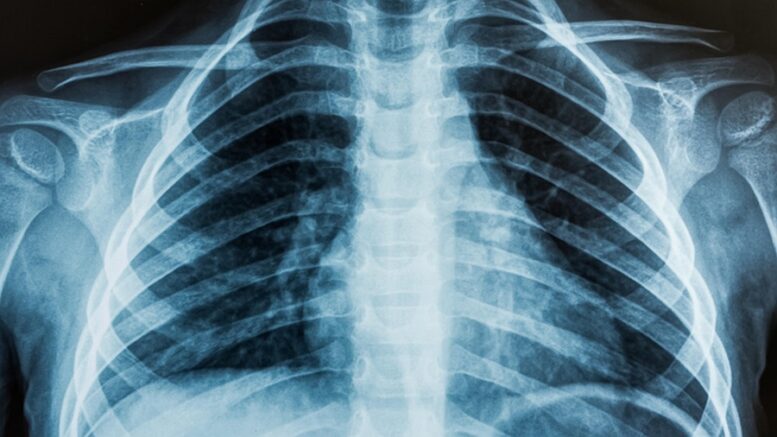
![]() Most of us are aware that X-ray imaging is incredibly useful for diagnosing injury and disease. But it’s also dangerous – ionizing radiation can damage DNA and cause cancer.
Most of us are aware that X-ray imaging is incredibly useful for diagnosing injury and disease. But it’s also dangerous – ionizing radiation can damage DNA and cause cancer.
The risk of X-rays can make headlines as much as the innovation. I worked on the development of X-ray imaging equipment for Pennsylvania-based industry bigwig DEXIS from 2011 to 2020, and just a few weeks before I joined the company, The New York Times published “Radiation Worries for Children in Dentists’ Chairs” – which generated lots of debate in our community and kept me quite busy for some time.
Of course X-rays are dangerous. But it’s important to note that we are exposed to ionizing radiation every day, mostly from energy that finds its way through our planetary atmosphere. In my opinion, that article was misleading – but I do like that it encouraged innovation that maintained diagnostic-image quality at the lowest possible radiation dose.
The equipment cited in the Times article – cone beam computed tomography – was invented in 1967 and developed for dental imaging in the 1990s. It enables the customized 3D reconstruction of a person’s anatomy for diagnostics and treatment planning. DEXIS (Imaging Sciences International, at the time) launched the dental-imaging system i-CAT in the mid-2000s and played a significant part in advancing the treatment of maxilla-facial disorders.

Brad Carlson: X factor.
In 2013, DEXIS launched an upgrade to the i-CAT – the i-CAT FLX, the first low-dose CBCT machine on the market. It decreased the amount of radiation used for some diagnostic images by two times or more, and for some scans the dose was no worse than walking down the street in Denver. Some scans required doses that were lower than a person receives during a transcontinental flight.
The go-to-market success of i-CAT was facilitated in large part by Melville-based Henry Schein, the largest global distributor of dental equipment and the exclusive U.S. distributor for i-CAT at the time.
So how do we know how much radiation we receive from X-ray machines, or from other sources? It starts with the amount of radiation produced by the source. X-rays are absorbed in our body differently by different kinds of tissue: Dense tissue, like bones, absorbs a lot of X-rays, while soft tissue absorbs less.
This is what creates the contrast in an X-ray image. Scientists have also developed models of human organs and tissue to account for the biological effect of radiation, measured in unites called sieverts.
On average, people receive about 5 uSv (microscopic doses) of background radiation every day. We’re exposed to more radiation at higher altitudes – as in, strolling through Denver or flying in a plane – and lower amounts at sea level.
The i-CAT FLX reduced the effective dose for some scans to the equivalent of a few days of background exposure, while providing image quality suitable for orthodontic and other dental treatments – improving medical diagnostics and treatment capabilities while decreasing patient risks.
Prior to the 1990s, X-ray images were produced with film that had to be processed chemically in a darkroom. During that decade, computed tomography was invented – film was replaced with photo-stimulated phosphor plates that could be digitized by specially designed machines.

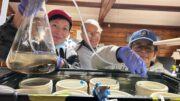
Moving things along: Aribex’s handheld Nomad device was a breakthrough in X-ray quality and safety.
In 2007, detectors were introduced that could directly absorb X-rays and produce digital images. These detectors paved the way for the design of medical computed tomography and CBCT.
Innovations have continued throughout this century. Consider that X-ray equipment long used a conventional vacuum tube that leveraged a high voltage (tens of kilovolts) to accelerate electrons onto a metal target, typically tungsten. The collision of electrons on the target produced X-rays, but in a highly uncontrolled way – they scattered in all directions. Only about 1 percent of the X-rays could be collimated into the cone directed at a target; the rest were blocked as much as possible, with the equipment operator often standing behind a shielded wall.
But in 2010, Utah-based Aribex developed a handheld X-ray source (the Nomad) that was safe enough for patients and operators alike. DEXIS parent KaVo Group, a German multinational, acquired Aribex in 2012 and continues to innovate the Nomad product.
Each advancement in detector technology has improved the detection efficiency reduced the required X-ray energy – and the risk to patients and operators. Today, efforts are underway to replace the traditional X-ray vacuum tube with modern technologies, including the use of carbon nanotubes and solid-state sources.
That’s good – continued innovation in X-ray imaging systems will continue to maximize benefits and reduce risks.
Brad Carlson is vice president of technology and business development at Hauppauge-based Intelligent Product Solutions.